Diatom, Colored scanning electron micrograph (SEM) - Stockillustratie
Diatom. Coloured scanning electron micrograph (SEM) of a diatom (Coscinodiscus sp.). Diatoms are a major group of eukaryotic algae, and are one of the most common types of phytoplankton. A characteristic feature of diatom cells is that they are encased within a unique cell wall made of silica (hydrated silicon dioxide), called a frustule. Magnification: x600 when printed at 10 centimetres wide.

Bestel uw ingelijste foto direct en bekijk de diverse opties op Photos.com.
LICENTIE KOPEN
Alle royalty free licenties bieden wereldwijde gebruiksrechten en uitgebreide bescherming. Daarnaast zijn er eenvoudige tarieven met volumekortingen beschikbaar.
€ 335,00
EUR
Getty ImagesDiatom Colored Scanning Electron Micrograph , Stockfoto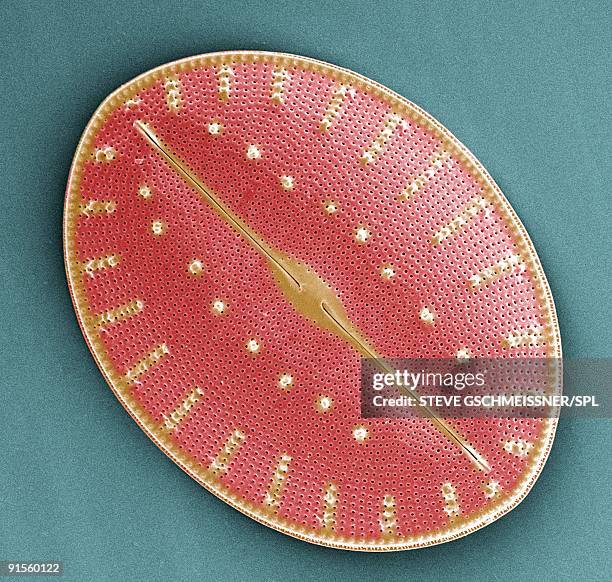 Download premium, authentieke Diatom, Colored scanning electron micrograph (SEM) stockillustraties van Getty Images. Verken vergelijkbare stockillustraties met hoge resolutie in onze uitgebreide visuele catalogus.Product #:91560122
Download premium, authentieke Diatom, Colored scanning electron micrograph (SEM) stockillustraties van Getty Images. Verken vergelijkbare stockillustraties met hoge resolutie in onze uitgebreide visuele catalogus.Product #:91560122€335€50
Getty Images
In stockGEGEVENS
Credits:
Creatief nr.:
91560122
Soort licentie:
Collectie:
Science Photo Library
Max. bestandsgrootte:
4319 x 4110 px (36,57 x 34,80 cm) - 300 dpi - 4 MB
Datum van uploaden:
Release-informatie:
Geen release vereist
Categorieën:
- Alg,
- Bescherming,
- Celmembraan,
- Clipart,
- Diatomee,
- Enkel object,
- Eukaryotic Cell,
- Fotosynthese,
- Frustule,
- Fytoplankton,
- Geel,
- Horizontaal,
- Illustratie,
- In het water levend organisme,
- Kleurcorrectie,
- Kleurenfoto,
- Microbiologie,
- Natuur,
- Ondersteuning,
- Plankton,
- Plant,
- Protozoa,
- Rasterelektronenmicroscoop,
- Rood,
- SEM,
- Uitsnede,
- Unicellular Organism,
- Wetenschap en Techniek,
- Zonder mensen,